- October 31, 2024
Echo Tech Enhancements: Glendale Memorial Hospital’s Commitment to Health in 2024

Understanding Echocardiography at Glendale Memorial Hospital: Key Techniques, Protocols, and Patient Care
Echocardiography, often called “echo,” is a crucial non-invasive method for examining the heart’s structure and function. At Glendale Memorial Hospital, echocardiography is integral to cardiac care, enabling physicians to detect, assess, and manage various heart conditions. Let’s explore the human side of this important diagnostic tool, from the imaging techniques used to the patient care protocols that ensure a positive experience.

1. Common Echocardiography Techniques Used
At Glendale Memorial Hospital, echocardiography involves various techniques tailored to each patient’s needs. The most frequently used method is transthoracic echocardiography (TTE), which is non-invasive and effective for visualizing the heart’s structure. For cases where TTE may not provide enough detail, transesophageal echocardiography (TEE) comes into play, offering high-resolution images of heart valves and posterior structures. Moreover, Doppler echocardiography, including color, pulse-wave, and continuous-wave Doppler, is utilized to assess blood flow and velocity through heart chambers and valves. Together, these methods offer a comprehensive understanding of heart health, assisting in diagnosing issues such as valve regurgitation or stenosis.
2. Patient Preparation Protocols
Preparing for an echocardiogram can vary depending on the type of procedure. For a standard TTE, patients often find it easy to fit into their routine, as they don’t need extensive preparation and can continue taking medications and eating. However, for a TEE, fasting for at least six hours before the procedure is necessary to minimize aspiration risks. Additionally, patients are encouraged to share information about their medications, especially blood thinners, which may require dosage adjustments. This preparation is essential for ensuring safety and achieving the best possible imaging results.
3. Ensuring Optimal Image Quality
Achieving clear and accurate images is a priority for the technicians at Glendale Memorial. They employ advanced positioning techniques and equipment settings to optimize image quality. For patients with challenging imaging conditions—like those who are overweight or have lung interference—contrast agents may be used to enhance the visibility of cardiac chambers. The hospital’s state-of-the-art ultrasound equipment allows adjustments in frequency and focus, which is especially critical in complex cases. These efforts reflect Glendale Memorial’s commitment to high diagnostic accuracy and effective patient care.
4. Calculating Ejection Fraction and Cardiac Function
At Glendale Memorial, the assessment of ejection fraction and cardiac function adheres closely to guidelines set by the American Society of Echocardiography (ASE) and the American Heart Association (AHA). The Simpson’s biplane method is the preferred technique for measuring ejection fraction, offering a precise evaluation of left ventricular function. For other cardiac parameters, ASE guidelines on chamber quantification are meticulously followed, ensuring that results are reliable and consistent, which in turn reassures patients about the accuracy of their diagnoses.
5. Doppler Settings and Flow Measurement Standards
Doppler echocardiography settings at Glendale Memorial are standardized to guarantee an accurate assessment of valvular function. Pulse-wave Doppler measures flow velocity at specific points, while continuous-wave Doppler evaluates high-velocity flows, which may indicate valve stenosis. Furthermore, color Doppler is instrumental in visualizing blood flow patterns, aiding in the detection of valvular regurgitation. By utilizing ASE-recommended parameters like peak and mean gradients for stenosis severity and vena contracta width for regurgitation quantification, the echocardiography team provides comprehensive insights into each patient’s cardiac health.
6. Safety in Contrast-Enhanced diographEchocary
Safety is a top priority at Glendale Memorial during contrast-enhanced echocardiography. The hospital conducts thorough screenings to identify any allergies to contrast agents and past adverse reactions. During the procedure, technicians remain vigilant, monitoring patients closely for any signs of discomfort or allergic reactions. Only FDA-approved contrast agents are used, and emergency protocols are in place to address any unforeseen reactions. Patients are informed and consenting, ensuring they feel secure throughout the process.
7. Reducing Radiation Exposure
While echocardiography typically doesn’t involve radiation, certain procedures that require fluoroscopic guidance, like TEE in specific interventional settings, may use radiation. Glendale Memorial emphasizes safety by utilizing low-dose fluoroscopy settings and providing protective equipment, such as lead aprons, for both patients and staff. The hospital follows the ALARA (As Low As Reasonably Achievable) principle, focusing on minimizing radiation exposure while still delivering quality diagnostic results.
8. Advanced Image Analysis and Reporting Tools
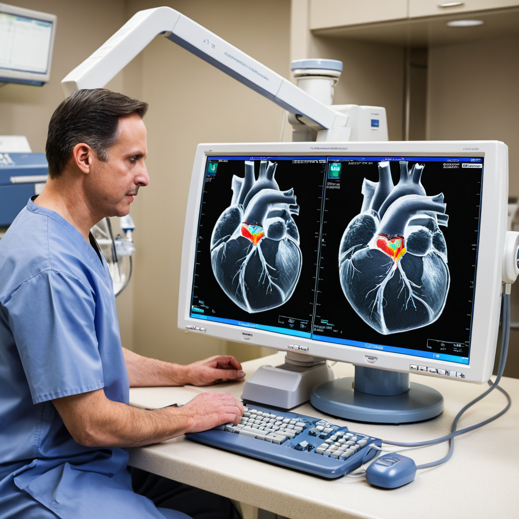
Glendale Memorial Hospital utilizes cutting-edge software like EchoPAC and Philips QLAB for post-procedure image analysis and reporting. These advanced tools enable comprehensive analyses, including 2D and 3D imaging, strain imaging, and precise quantification of cardiac structures and function. Moreover, the software integrates smoothly with the hospital’s electronic health record (EHR) system, allowing physicians to access images and reports efficiently. This integration enhances patient care and supports timely decision-making.
9. Routine Maintenance of Equipment
To maintain accuracy and prevent technical issues, Glendale Memorial ensures regular maintenance of its echocardiography equipment. Routine maintenance is conducted every six months according to manufacturer recommendations and hospital policies. Daily quality control checks are also performed by technical staff to ensure that all components, such as probes and transducers, are functioning optimally. If any malfunctions are detected, certified technicians undertake immediate repairs, preserving the integrity of diagnostic results.
10. Handling Unexpected Findings
In cases of unexpected findings, such as new valve abnormalities or signs of ventricular dysfunction, Glendale Memorial has established protocols to ensure prompt follow-up. Echocardiography technicians collaborate closely with supervising cardiologists for further evaluation. If the findings are critical, the cardiologist may discuss them with the patient immediately, ensuring that timely intervention can occur. Additionally, thorough documentation of results is shared with the patient’s primary care or treating physician to facilitate a coordinated approach to care.
Conclusion
Echocardiography at Glendale Memorial Hospital goes beyond just advanced technology; it embodies a comprehensive approach that prioritizes patient safety, skilled professionals, and effective communication. By adhering to strict guidelines, employing state-of-the-art imaging tools, and focusing on patient-centered care, Glendale Memorial ensures that every echocardiography procedure provides valuable insights into patient health, leading to accurate diagnoses and improved treatment outcomes. The dedicated team is committed to making the process as comfortable and informative as possible, helping patients navigate their cardiac health with confidence.